
The production of imaging apparatus for observation of the distribution of medicine in the body of animal animals by Iranian researchers.
June 17, 2019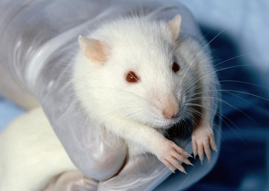
Laboratory animals are not sacrificed
If we use many of the medicines and treatments that have created in labs, this should be owed to the mice and other creatures who, during the laboratory and to ensure the correct functioning of the process, dangerous and sometimes deadly tests first they are done on them.
Currently, for testing the new drug, the animal is killed after the injection of drug and the tissue is cut from the animal; the effects of the drug on the patient are examinedafter the pathology. However, using a molecular fluorescence tomography system, without the need for tissue cutting, it can be performed more effectively and identify many diseases, including cancer, at the very beginning of the formation.
DrMarjanehHejazi, a speaker of the design of a molecular fluorescent cutting system, has talked about the benefits of this method and the possibilities that the device provides to researchers.
What are the advantages of what you do and use fluorescence for this tomography?
Tomography is a method for early diagnosis of cancer at the cellular level. The advantages of this method are non-invasive, easy to use, the use of non-ionizing radiation and the detection of cellular lesions.
When did the initial idea of the production of this machine come from and at what stage?
The initial idea of this machine was started from student projects conducted at Tehran University of Medical Sciences. Then, through several projects with related groups and obtaining appropriate financial resources from the competent authorities, they reached the stage of commercialization. I and my colleagues at TajhizAfarinanNooriParseh, affiliated with Tehran University of Medical Sciences, managed to construct a molecular fluorescence imaging system for small animals. The device is currently ready for use in research centers.
Generally, fluorescent imaging is a new method that has been commercially used by medical and research centers abroad for about three or four years. The fluorescent molecular method is currently used in the United States and most European countries.
Is the use of molecular imaging limited to preclinical and research applications?
The system is widely used in small animal imaging in the field of Nano drugs, in the cancer treatments and in the diagnosis of early stage cancer, and is now widely used in the pharmaceutical industry to examine the properties of drugs and Nano drugs. Of course, this device is used for pre-clinical and research stages.
How can this system be used in the research and treatment of various diseases?
Using this device leads to a more detailed study of the drugs and the follow-up of the effects of new therapies on the small animal. Then, with this study, it is possible to generalize the results to mankind. In this way, the cost of treatment is reduced and the accuracy of diagnostic and therapeutic methods is greatly increased.
What is the diagnostic error in this method and how much this error can affect the results of the tests? What should be done to reduce the error rate?
Hejazi: The molecular fluorescence tomography method is a method for early diagnosis of cancer at the cellular level, which will definitely be among the early needs of pharmaceutical industries and related research centers in the near future.
In this machine, the nanoparticles are concentrated in the target tissues, so this method is highly accurate. In order to improve the accuracy of the diagnosis, making the nanoparticles suitable for accumulation in the waste is very important.
How does this device work?
In the usual way after the injection of the nanoparticle into the animal, the animal is killed and sampling is done.
Samples prepared from the target tissue are sent to the relevant diagnostic centers. In this way, the possibility of evaluating the effects in living tissue is eliminated. In the molecular fluorescence method, the small animal during the study is only anesthetized, so the study is performed under normal conditions.
What are the components of molecular fluorescence cutting system, and what kind of operation can each part provide for the system?
This machine consists of three lasers, a very sensitive camera, and a mechanical and electronicstructure suitable for shooting 360 degrees of a small animal. In addition, this machine includes software written by experts inside the country that allows the investigation of the three-dimensional distribution of fluorescent material in the target lesion.
Laser beams also allow for a wide range of nanoparticles, organic fluorescent materials, cells and labeled tissues. In addition, the unique mechanical and electronic structure of this machine makes it possible to examine the body parts of the animal in different ways.
How do you see the future of these systems and what other applications do you expect them to have?
Considering that the molecular fluorescence method in the world is very new and has recently been widely used in various medical fields, it will definitely be among the early needs of pharmaceutical industries and related research centers in the near future. At present, the device is designed to evaluate the function of cells and tissues.
The research team is researching the device to add anatomical information to the functional information of small animal tissues. Therefore, in the near future, it is possible to detect lesions in their anatomical location.

